

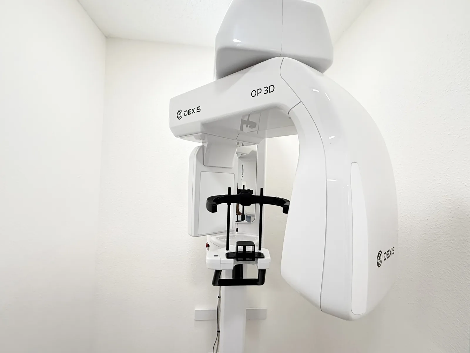
What is 3D CBCT Dental Imaging?
Cone Beam Computed Tomography (CBCT) is an advanced medical-grade imaging technology that emits a cone-shaped X-ray beam around a patient's head to produce hundreds of individual high-resolution 2D planar images. Specialized computer software compiles these cross-sections into a complete, distortion-free three-dimensional volumetric digital model of your teeth, roots, maxillary sinuses, mandibular nerve canals, and alveolar jawbone.
Unlike conventional 2D dental X-rays (such as bitewings or panoramic films) that flatten complex three-dimensional anatomy into a single distorted overlay, CBCT allows our clinical team to inspect anatomical structures from any angle, measure bone density down to fractions of a millimeter, and plan complex surgical procedures with absolute predictability.
Why 3D CBCT Outperforms Traditional 2D Dental X-Rays
For more than a century, dentistry relied entirely on two-dimensional radiographic film. While 2D X-rays remain useful for detecting simple surface cavities between teeth, they present significant diagnostic blind spots for complex specialty procedures:
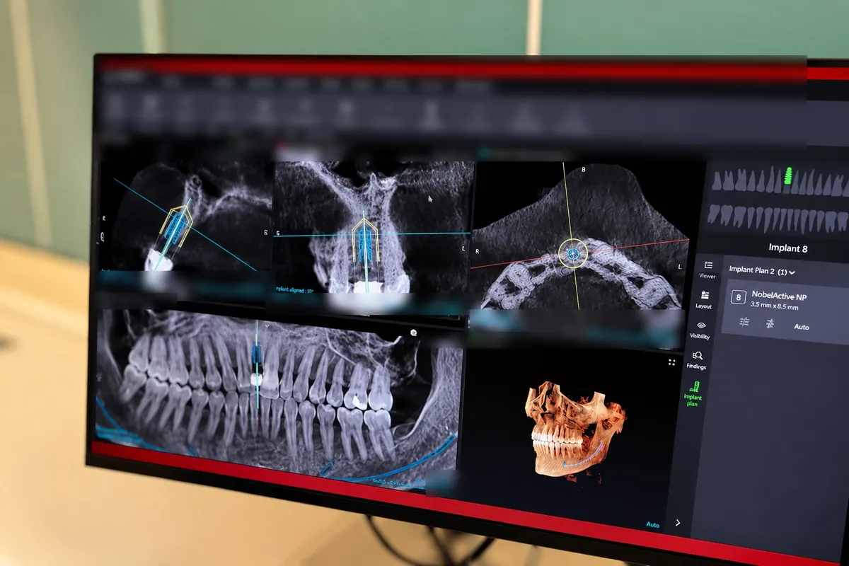
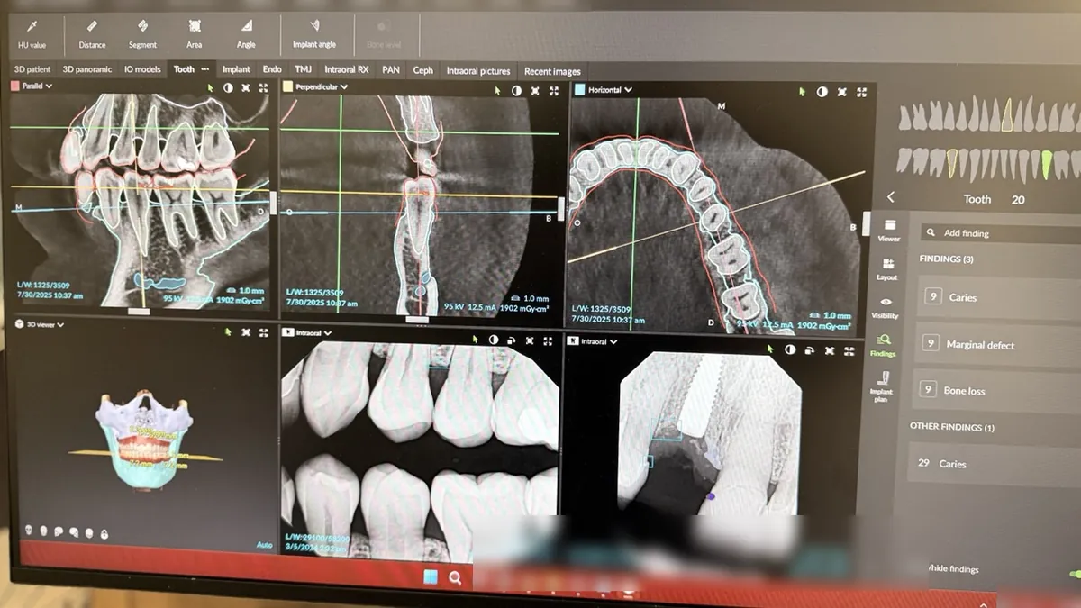
- Elimination of Anatomical Superimposition: On a standard 2D panoramic X-ray, the dense cortical bone of the cheekbone (zygoma), spinal column, and opposite jaw are superimposed on top of your teeth, hiding root fractures, cysts, and subtle bone defects. CBCT slices through overlapping layers with zero distortion.
- True 1:1 Scale Measurement: Traditional panoramic X-rays introduce 15% to 25% geometric magnification and elongation. A 3D CBCT scan delivers exact 1:1 true physical dimensions, ensuring that an implant planned for 10.5mm is placed with micrometer accuracy.
- Bone Density Quantification: CBCT calculates bone quality (cortical thickness and trabecular density) in Hounsfield-equivalent units, allowing our implant dentists to determine whether immediate loading (such as All-on-4 same-day teeth) is biologically viable.
- Nerve & Sinus Safety: The inferior alveolar nerve canal running through your lower jaw is clearly mapped in 3D cross-section, eliminating the risk of nerve impingement or permanent numbness during dental implant placement or wisdom tooth extraction.
Radiation Safety: The ALARA Principle & Dose Comparison
Patient safety is paramount at Aloha Dental Specialty Center. We strictly adhere to the ALARA principle (As Low As Reasonably Achievable). Our modern CBCT machine utilizes pulsed cone beam technology, meaning the X-ray tube emits radiation only in short microsecond bursts during its rotational path, rather than continuous exposure.
To put radiation exposure into clear medical perspective, consider how dental CBCT compares to everyday environmental radiation and medical hospital CT scanners:
| Radiation Source | Estimated Effective Dose (µSv) | Equivalent Natural Background Radiation |
|---|---|---|
| Small Field-of-View Dental CBCT (Endodontic) | 15 – 35 µSv | Approx. 2 to 4 days of normal background life |
| Medium Field-of-View CBCT (Single Jaw / Implants) | 30 – 80 µSv | Approx. 4 to 10 days of normal background life |
| Standard Full-Mouth 2D X-Ray Series (FMX) | 35 – 85 µSv | Approx. 5 to 11 days of background radiation |
| Cross-Country Commercial Flight (Roundtrip NY-LA) | 40 µSv | Approx. 5 days of atmospheric radiation |
| Average Natural Background Radiation (Per Year) | 3,000 µSv | Normal background radiation from sun, soil, rocks |
| Hospital Medical Head CT (Non-Dental) | 1,500 – 2,500 µSv | Approx. 6 to 10 months of background radiation |
As the data demonstrates, a dental CBCT scan delivers up to 90% less radiation than a conventional hospital medical CT, providing maximum diagnostic clarity with minimal radiation exposure.
Clinical Applications Across Specialties at ADSC
Our in-house 3D CBCT scanner serves as the diagnostic cornerstone across all dental specialty disciplines at Aloha Dental Specialty Center:
3D Guided Dental Implants
Dr. Merat Ostovar uses 3D CBCT scans to virtually plan implant positioning, angle, and depth within specialized CAD software before 3D-printing custom surgical guides for millimeter-precise placement.
Endodontic Root Canal Precision
Our endodontic specialists utilize targeted small-FOV CBCT scans to uncover hidden canals (such as the elusive MB2 canal), evaluate vertical root fractures, and diagnose persistent tooth pain.
Impacted Wisdom Teeth Removal
3D cross-sectional views reveal the exact relationship between impacted wisdom tooth roots and the inferior alveolar nerve, virtually eliminating the risk of temporary or permanent nerve paresthesia.
Periodontal Bone Architecture
Periodontist Dr. Walied Eldomiaty evaluates three-dimensional infrabony defects, furcation involvement between molar roots, and tracks bone regeneration following LANAP laser therapy.
Airway & Sleep Apnea Analysis
CBCT software computes total pharyngeal airway volume and identifies minimum cross-sectional airway constriction, assisting in the design of custom oral sleep appliances.
TMJ Condylar Diagnostics
High-resolution imaging of the temporomandibular joints reveals condylar erosion, flattening, osteophytes (bone spurs), and joint space asymmetries causing jaw clicking or pain.
What to Expect During Your 14-Second 3D Scan
If you have ever felt claustrophobic inside a hospital MRI or CT scanner tube, you will be relieved by the open, comfortable design of our dental CBCT unit:
- Open-Air Standing or Seated Position: You simply stand or sit comfortably upright in an open room. There are no enclosed tunnels, zero claustrophobia, and no feeling of confinement.
- Simple Head Alignment: You rest your chin on a gentle, sanitized support pad and bite lightly on a small disposable plastic wafer. Two soft temple supports stabilize your head to prevent movement.
- Quick 14-Second Rotation: The C-arm of the scanner smoothly rotates 360 degrees around your head. The scan takes approximately 14 seconds and is completely silent and painless.
- Immediate 3D Chairside Review: Within seconds, your 3D digital model renders on our chairside monitors. Your doctor reviews your anatomy with you in real time, rotating your jaw in 3D space to explain findings clearly.
Scan-Only Services for Referring Dentists
Aloha Dental Specialty Center proudly serves as a 3D imaging resource for general dentists and dental specialists throughout Beaverton, Aloha, Hillsboro, and the greater Portland metro area who do not have an in-house CBCT machine:
- Rapid Turnaround: Referring offices can send patients for a convenient scan-only appointment, typically completed in under 15 minutes.
- Full DICOM Export: Scan files are exported in standard multi-slice DICOM format onto secure USB media or transmitted via encrypted HIPAA-compliant cloud portals.
- Integrated 3D Viewer Software: Every scan export includes complimentary 3D slice viewing software, allowing referring doctors to manipulate cross-sectional views, perform nerve tracing, and measure bone dimensions in their own offices.
- Prompt Patient Return: We strictly perform the requested imaging and return the patient directly to your office for continued treatment. Referrals can be submitted via our referring doctor portal.
Insurance Billing & Cost for Dental CBCT
The cost of a dental CBCT scan typically ranges from $250 to $450 depending on the field-of-view (focused local scan vs. full dual-arch maxillofacial scan). When performed as part of surgical planning for dental implants, bone grafting, endodontic retreatment, or wisdom teeth removal at ADSC, the imaging fee is factored into your comprehensive treatment plan.
Insurance Coverage: Many dental PPO plans provide coverage for CBCT scans under diagnostic codes CDT D0364 (cone beam CT capture and interpretation for one jaw) or CDT D0367 (both jaws). In cases involving impacted teeth, severe infections, or sleep apnea, medical health insurance may also contribute to diagnostic imaging costs. We verify all available benefits on your behalf prior to imaging.
Frequently Asked Questions About 3D CBCT Imaging
Yes, dental CBCT imaging is exceptionally safe. Because it uses pulsed cone-beam technology targeted specifically to the oral cavity, the radiation dose is up to 90% lower than medical hospital CT scans. A focused dental scan emits approximately 20 to 50 micro-sieverts (µSv), equivalent to just a few days of normal environmental background radiation or a roundtrip cross-country airline flight.
Not at all. Unlike hospital CT or MRI machines where you lie inside an enclosed, narrow tube, our dental CBCT unit is completely open. You stand or sit comfortably upright in an open room. The scanner arm simply rotates around the outside of your head for 14 seconds without touching you.
Regular dental X-rays provide only a flat, two-dimensional picture with distorted, overlapping shadows. They cannot show bone width, bone density, the 3D position of nerves, or complex root canal curves. For procedures like dental implants, wisdom teeth extraction, bone grafting, and root canals, a 3D CBCT scan is essential to prevent nerve injury, avoid sinus perforation, and ensure surgical success.
Yes. We provide complete DICOM files along with built-in 3D viewer software on a flash drive or via secure electronic transfer for any referring doctor, physician, or specialist upon request.
Many dental PPO insurance plans provide coverage for 3D CBCT imaging under codes D0364 or D0367 when deemed medically necessary for surgical treatment planning, such as impacted teeth, implant surgery, or endodontic pathology. Our insurance team checks your specific benefits and submits claims on your behalf.